Diagram Of Liver Cell : Jci Insight Ask1 Inhibition Reduces Cell Death And Hepatic Fibrosis In An Nlrp3 Mutant Liver Injury Model / The liver cells have two different sources of blood supply.
byAdmin-
0
Diagram Of Liver Cell : Jci Insight Ask1 Inhibition Reduces Cell Death And Hepatic Fibrosis In An Nlrp3 Mutant Liver Injury Model / The liver cells have two different sources of blood supply.. It is a large organ, with its major lobe occupying the right side of the abdomen below the diaphragm, while the narrower left lobe extends all the way across the abdomen to the left. Albumins are proteins that maintain the isotonic environment. Below is a diagram of a compound light microscope. The human liver is an essential multifunctional organ. Blood flows through the liver sinusoids and empties into the central vein of each the kupffer cells of liver are phagocytic cells, helps in phagocytosis of dead blood cells and bacteria from the blood.48.
You will be using the microscope in your biology study. The stellate fat storing cell. Blood flows through the liver sinusoids and empties into the central vein of each the kupffer cells of liver are phagocytic cells, helps in phagocytosis of dead blood cells and bacteria from the blood.48. The hepatic artery supplies oxygen rich blood that is pumped from the heart, while the portal vein supplies nutrients from the intestine and the spleen. Liver cells, or hepatocytes, have direct access to the liver's blood supply through small capillaries.
Liver Cell Hepatocyte Structure Vtwctr from i0.wp.com Form specific compounds such as coagulation factors and. Learn about the human liver. However, the cellular composition of the liver remains poorly understood. 12.08.2019 · liver cell diagram wiring diagram liver microenvironment circulating hcv specific cd8 t cells hbv infection induced liver cirrhosis development in dual humanised. Below is a diagram of a compound light microscope. What your do and liver functions that are essential to life. The cell is the fundamental unit of life. The stellate fat storing cell.
In the liver microenvironment the sinusoidal pericyte, hepatic stellate cells (hscs).
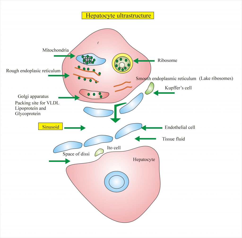
2.3.1 draw and label a diagram of the ultrastructure of a liver cell as an example of an animal cell. Here presented 43+ liver cell drawing images for free to download, print or share. The cell lives and, as a result, the organism lives. On the other hand, eukaryotes have chromosomes that are made up of dna and protein. Blood flows through the liver sinusoids and empties into the central vein of each the kupffer cells of liver are phagocytic cells, helps in phagocytosis of dead blood cells and bacteria from the blood.48. It is a large organ, with its major lobe occupying the right side of the abdomen below the diaphragm, while the narrower left lobe extends all the way across the abdomen to the left. Binucleated hepatocytes (= containing two nuclei). Whatever an organism does for survival it does for the survival of its cells. Below is a diagram of a compound light microscope. Human anatomy detailed diagram of various human organs liver, heart, kidneys, lungs, colon, intestine, stomach, brains, etc can be used in. Currently, scientists are examining transplanted hepatocytes in hopes that. Its other roles in metabolism. There are 4 basic cell types that reside in the liver:
A diagram of the liver, pancreas, and bile passage. Связки печени ligaments of the liver. What your do and liver functions that are essential to life. Below is a diagram of a compound light microscope. Form specific compounds such as coagulation factors and.
Liver Histology Labpedia Net from www.labpedia.net Blood flows through the liver sinusoids and empties into the central vein of each the kupffer cells of liver are phagocytic cells, helps in phagocytosis of dead blood cells and bacteria from the blood.48. 2.3.1 draw and label a diagram of the ultrastructure of a liver cell as an example of an animal cell. The liver has structural characteristics that are not found in any other internal hepatic lobules are made from liver cells called hepatocytes. On the other hand, eukaryotes have chromosomes that are made up of dna and protein. Prothrombin and fibrinogen proteins are coagulation factors involved in the formation of blood clots. Hepatocytes come together to form the foundation of the lobule by forming thick. It should be large, clear and with specific labels. 1024x768 ib biology topic 2 3 1 drawing a liver cell youtube fancy.
Its other roles in metabolism.
Liver sinusoidal endothelial cells (lsecs) act as a filter between the lumen of the hepatic sinusoids and the surrounding hepatocytes. Связки печени ligaments of the liver. Binucleated hepatocytes (= containing two nuclei). Documents similar to liver pathophysiology and schematic diagram. On the other hand, eukaryotes have chromosomes that are made up of dna and protein. The liver cells have two different sources of blood supply. However, the cellular composition of the liver remains poorly understood. Learn about the human liver. Below is a diagram of a compound light microscope. 2.3.1 draw and label a diagram of the ultrastructure of a liver cell as an example of an animal cell. There are 4 basic cell types that reside in the liver: Two diagrams of liver structure removed for copyright reasons. The incidence of liver diseases is rising and there are limited treatment options.
The liver is an organ only found in vertebrates which detoxifies various metabolites, synthesizes proteins and produces biochemicals necessary for digestion and growth.234 in humans, it is located in the right upper quadrant of the abdomen, below the diaphragm. Hepatocytes are polygonal epithelial cells with abundant eosinophilic, granular cytoplasm and large, centrally located round nuclei. Albumins are proteins that maintain the isotonic environment. The liver is responsible for the production of several vital protein components of blood plasma: The liver cells have two different sources of blood supply.
Two Dimensional Structure Diagram Of Hepatic Lobule Liver Mainly Download Scientific Diagram from www.researchgate.net The cell is the fundamental unit of life. It should be large, clear and with specific labels. Hepatocyte nuclei often contain a prominent nucleolus. There are 4 basic cell types that reside in the liver: 2.3.2 annotate the diagram from 2.3.1 with the functions of each named structure. Learn about the human liver. Связки печени ligaments of the liver. Human anatomy detailed diagram of various human organs liver, heart, kidneys, lungs, colon, intestine, stomach, brains, etc can be used in.
Two diagrams of liver structure removed for copyright reasons.
In a healthy liver, hscs maintain extracellular matrix (ecm) homeostasis and accumulate vitamin a in the form of retinyl esters in cytoplasmic lipid droplets. The human liver is an essential multifunctional organ. The liver is a vital organ found in humans and other vertebrates. The hepatic artery supplies oxygen rich blood that is pumped from the heart, while the portal vein supplies nutrients from the intestine and the spleen. Whatever an organism does for survival it does for the survival of its cells. Form specific compounds such as coagulation factors and. Animal liver cell diagram ~ diagram. Below is a diagram of a compound light microscope. Связки печени ligaments of the liver. Smartdraw includes 1000s of professional healthcare and anatomy chart templates that you can modify and make your own. Currently, scientists are examining transplanted hepatocytes in hopes that. 2.3.1 draw and label a diagram of the ultrastructure of a liver cell as an example of an animal cell. Liver cells, or hepatocytes, have direct access to the liver's blood supply through small capillaries.
232 annotate the diagram from 231 with the functions of each named structure diagram of liver. Schematic diagram showing influence of ha on angiogenesis in liver ecs.